tree in bud opacities pneumonia
As in this case renal cell carcinoma is one of the most common malignancies that may produce this vascular cause of tree-in-bud pattern. More extensive lympho - cytic infiltrations may be associated with lymphoid interstitial pneumonia LIP with ground-.

High Resolution Computed Tomography Patterns Of Diffuse Interstitial Lung Disease With Clinical And Pathological Correlation Younger Survival Pulmonar
Mycobacterium avium complex is the most common cause in most series.

. Studies have reported that pulmonary TB accounts for only 28 of the cause of tree-in-bud opacities as opposed to pulmonary apical granulomas and fibrosis being more suspicious of. A young male patient who had a history of fever cough and respiratory distress presented in the emergency department. 1 direct filling of the centrilobular arteries by tumor emboli and 2 fibrocellular intimal hyperplasia due to carcinomatous endarteritis.
Note the scattered lung nodules surrounded by. Simply put the tree-in-bud pattern can be seen with two main sites of disease 3. Patients with normal standard physiological pulmonary tests have been shown to have mosaic perfusion and air trapping on HRCT suggestive of bronchiolitis obliterans and a pattern of branching linear opacities like a tree in bud appearance suggestive of bronchiectasis with mucoid secretions.
Subsequently question is what causes tree in bud opacities. However gram staining and cultures were negative. Chest radiography had demonstrated signs of bronchiectasis and several scattered nodules Figure 2.
1 It is important for clinicians to remember that this pattern has an extensive. Although initially described in 1993 as a thin-section chest CT finding in active tuberculosis TIB opacities are by. Distal pulmonary vasculature More specifically the pattern can be manifest becaus.
Classically bronchiolitis appears as a region of centrilobular nodularity often in a tree-in-bud pattern. Pneumonia due to respiratory syncytial virus in a 23-year-old man with leukemia. The purpose of this study was to determine the relative frequency of causes of TIB opacities and identify patterns of disease associated with TIB opacities.
3 Aspiration is also a common cause of the tree-in-bud formation. 1012 Poorly defined centrilobular nodules associated with branching linear and nodular opacities ie tree-in-bud sign are the typical HRCT findings of infective bronchiolitis frequently. These small clustered branching and nodular opacities represent terminal airway mucous impaction with adjacent peribronchiolar inflammation.
The patient underwent CT scanning of the chest which showed areas of nodular infiltration in the lower lobes with tree. In radiology the tree-in-bud sign is a finding on a CT scan that indicates some degree of airway obstruction. In the acute phase bacterial pneumonia manifests in the form of segmental or lobar consolidation Fig 2 possibly with cavitation and related hilar and mediastinal adenopathies.
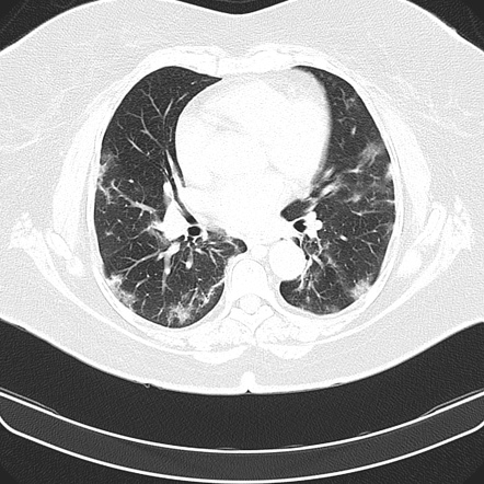
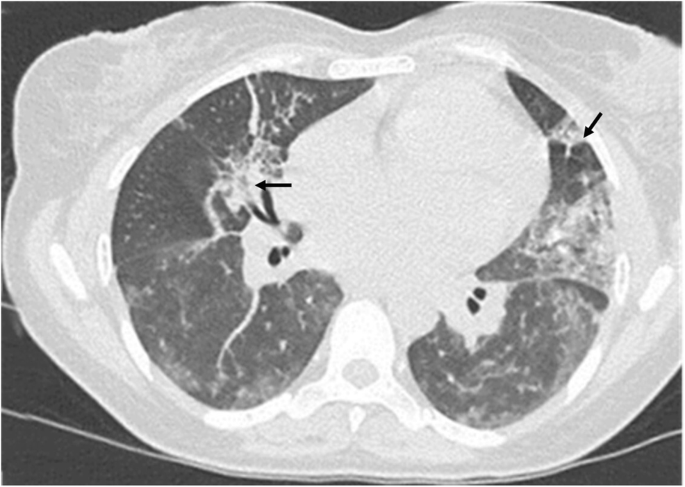
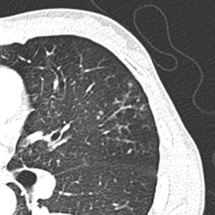
However to our knowledge the relative frequencies of the causes have not been evaluated. Thin-section CT scan shows peripheral poorly defined centrilobular nodules and tree-in-bud opacities bilaterally. Malignancy can be associated with the tree-in-bud sign.
Usually somewhat nodular in appearance the tree-in-bud pattern is generally most pronounced in the lung periphery and associated with abnormalities of the larger airways. Aspiration pneumonia also mostly involves lower lobes and the posterior lung and can manifest as patchy GGOs andor consolidations. Tree-In-Bud Pattern A lymphoid interstitial infiltrate in the walls of the small airways follicular bronchiolitis may cause small centrilobular nodules and the tree-in-bud pattern Fig.
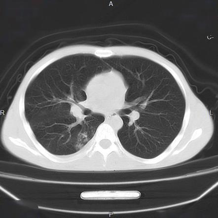
A chest radiograph showed bilateral nodular opacities with a left lower lobar consolidative opacity Fig 1A 1B. Tree-in-bud refers to a pattern seen on thin-section chest CT in which centrilobular bronchial dilatation and filling by mucus pus or fluid resembles a budding tree. It is most commonly associated with infectious diseases affecting the bronchioles1 OP resulting in a tree in bud pattern has been previously suggested2 However a clear radiological-pathological correlation of OP filling the bronchioles resulting in a tree in bud pattern has to the best of our knowledge not yet been clearly demonstrated.
Patients with aspiration pneumonia are some-times complicated with Mycobacterium infections especially elderly patients. And tree-in-bud branching opacities detected throughout both lung fields after aspiration. Tree-in-bud TIB opacities are a common imaging finding on thoracic CT scan.
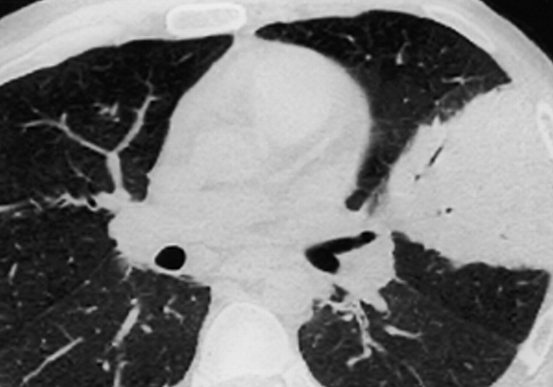
TIB opacities represent a normally invisible branches of the bronchiole tree 1 mm in diameter that are severely impacted with mucous pus or fluid with resultant dilatation and budding of the terminal bronchioles 2 mm in diameter1 photo. A tree-in-bud pattern of centrilobular nodules from metastatic disease occurs by two mechanisms. Vealed scattered linear nodular and tree-in-bud opacities involving the bilateral apices and the upper middle and lower lobes of the right lung suggestive of bronchiolitis.
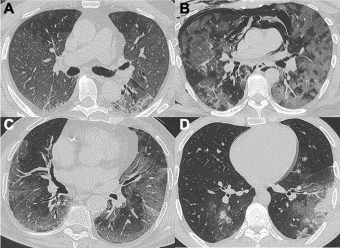
Associated focal ground-glass and consolidative opacities may be visualized although this should not the predominant feature. We here describe an unusual cause of TIB during the COVID-19 pandemic. The purpose of this study was to determine the relative frequency of causes of TIB opacities and identify patterns of disease associated with TIB opacities.
Patients with aspiration pneumonia are some-times complicated with. Tree-in-bud TIB appearance in computed tomography CT chest is most commonly a manifestation of infection. Organizing pneumonia most commonly results in a patchy bilateral consolidation that has a.
In radiology the tree-in-bud sign is a finding on a CT scan that indicates some degree of airway obstruction. Nodular opacities with tree-in-bud appearance can be associated with other changes in lung parenchyma-such as thickening of the bronchial walls consolidations andor areas of. Adjacent bronchial wall thickening is also frequently depicted.
Distal airways more common 2. HR-CT patterns seen in OP are. Mild bronchiectasis had also been noted Figure 1.
Multiple causes for tree-in-bud TIB opacities have been reported. The tree-in-bud sign is a nonspecific imaging finding that implies impaction within bronchioles the smallest airway passages in the lung. Tree-in-bud TIB opacities are a common imaging finding on thoracic CT scan.
The differential for this finding includes malignant and inflammatory. While the findings of bronchiolitis such as centrilobular nodular opacities and a tree-in-bud pattern are common in aspiration pneumonia they are not typically found in COVID-19 pneumonia 61 62 Fig. 2 However the classic cause of tree-in-bud is Mycobacterium tuberculosis especially when it is active and contagious and associated with cavitary lesions.
Ground Glass Opacity Ggo A Review Of The Differential Diagnosis In The Era Of Covid 19 Springerlink

Pin By Saikat Bhattacharjee On Radio Gaga Radiology Medicine Radio
Covid 19 Pneumonia Radiology Case Radiopaedia Org
Imaging Of Pneumonia Trends And Algorithms European Respiratory Society

Round Pneumonia Fish Pet Pets Pneumonia

Pin De Saikat Bhattacharjee En Radiology
Tree In Bud Sign Lung Radiology Reference Article Radiopaedia Org
Chest Ct Mimics Of Covid 19 Pneumonia A Review Article Springerlink
2

Tree In Bud Sign Lung Radiology Reference Article Radiopaedia Org

Pneumocystis Jirovecii Pneumonia At Chest High Resolution Computed Tomography Hrct In Non Hiv Immunocompromised Patients Spectrum Of Findings And Mimickers European Journal Of Radiology

Multimodality Imaging Of Covid 19 Pneumonia From Diagnosis To Follow Up A Comprehensive Review European Journal Of Radiology

Cavity Consolidation With Multiple Areas Of Nodular Opacity Showing Tree In Bud Appearance Most Likely Possibility Of Character Fictional Characters Snoopy

Tree In Bud Sign An Overview Sciencedirect Topics

Cavity Consolidation With Multiple Areas Of Nodular Opacity Showing Tree In Bud Appearance Most Likely Possibility Of E Opacity Abstract Artwork Abstract
Tree In Bud Sign An Overview Sciencedirect Topics

Review Of Chest Ct Manifestations Of Covid 19 Infection European Journal Of Radiology Open
Tree In Bud Sign Lung Radiology Reference Article Radiopaedia Org

24 Year Old Man With M Pneumoniae Pneumonia Ct Shows Centrilobular Download Scientific Diagram